Pracownie naukowe

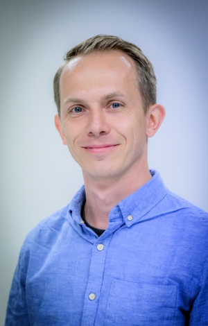
Dr hab. Michał Turek
Pracownia Fizjologii Molekularnej ZwierzątZakres badań
A new proteostasis mechanism for the removal of protein aggregates and damaged mitochondria has recently been described. Within this process, cellular waste material is ejected from the cell via large vesicles, termed exophers. Our laboratory is interested in deciphering the regulation of muscular exopher formation and uncovering its additional roles that go beyond maintaining protein/organelle homeostasis.
Badania
Najważniejsze osiągnięcia badawcze
- We identified muscular exophers in Caenorhabditis elegans as a model organism.
- We found that exopher production in C. elegans is a non-cell autonomous process regulated by egg formation in the uterus, which is used to nourish and improve the growth rate of the next generation.
- We discovered that social interactions and environmental cues significantly influence exopher production through complex pheromone-based communication mechanisms.
Opis badań
To maintain proper cellular protein homeostasis, the tight regulation of protein synthesis, protein degradation, and the subtle balance between these two processes must be maintained. New proteins are made on ribosomes, and unnecessary or damaged proteins are recycled predominantly by the ubiquitin-proteasome system and autophagy-lysosome pathway. Recently, a complementary proteostasis mechanism has been described in Caenorhabditis elegans neurons and murine cardiomyocytes. Under proteotoxic stress, neurons in worms can remove protein aggregates, damaged mitochondria, and the lysosome into neighboring tissues via large membrane-surrounded vesicles called exophers. Exophers are much larger than previously described vesicles (approximately 4 µm in diameter), and their formation enables neurons to maintain their proper functionality during neurotoxic stress and aging (Melentijevic et al. 2017). Moreover, the ejection of dysfunctional mitochondria from cardiomyocytes by exophers was recently reported (Nicolas-Avila et al. 2020). The evolutionary conservation of this cellular material extrusion phenomenon suggests that it constitutes a significant but currently poorly understood pathway that serves to maintain protein/organelle homeostasis. Indeed, using specially designed fluorescent reporters, we observed that C. elegans’ body wall muscles also produce high levels of exophers that are larger (up to 20 µm) than neuronal variants.
Using worms that express fluorescent reporters in body wall muscle cells, we found that exopher formation (exopheresis) is a non-cell autonomous process that is regulated by egg formation in the uterus. Our data suggest that exophers serve as transporters for muscle-generated yolk proteins that are used to nourish and improve the growth rate of the next generation. We propose that one of the roles of muscular exopheresis is to stimulate reproductive capacity, thereby influencing the adaptation of worm populations to current environmental conditions (Turek et al. 2020). These data formed the basis for our SONATA grant application, allowing us to continue this project as the main focus of our research group in the coming years.
Our recent findings emphasize the significant role of pheromone-based communication in exophergenesis. Social interactions and environmental cues have been shown to influence exopher production, revealing a complex regulatory network involving ascarosides, G protein-coupled receptors (GPCRs), and olfactory neurons. This network dynamically regulates muscle extracellular vesicle production in response to environmental signals (Szczepańska et al. 2024).
Despite research suggesting that exophers may be a fundamental mechanism for regulating cellular function, many aspects of this process remain unclear. Moving forward, our research will focus on the following key questions:
- What is the mechanism of exopheresis regulation?
- How does environmental information influence exopher generation?
- What role does the nervous system play in regulating muscular exopheresis?
- How does epigenetic regulation affect exopheresis, and what impact might it have on transgenerational inheritance?
Bibliography
- Melentijevic et al. Nature 2017. doi: 10.1038/nature21362
- Nicolas-Avila et al. Cell. 2020. doi: 10.1016/j.cell.2020.08.031
- Szczepańska et al. Nature Communications. 2024. doi: 10.1038/s41467-024-47016-x
- Turek et al. BioRxiv. 2020. doi: 10.1101/2020.06.17.157230
Metodologia
Brightfield microscopy, epi-fluorescent microscopy, confocal microscopy, high-throughput microscopic screens, optogenetics, calcium imaging, CRISPR/Cas9, transcriptomics, proteomics, behavioral assays, lifespan measurements.
Wybrane publikacje
- Pheromone-dependent olfaction bidirectionally regulates muscle extracellular vesicles formation. Banasiak K, Szczepańska A, Kołodziejska K, Ibrahim AT, Pokrzywa W*, Turek M*. bioRxiv preprint. 2022. doi: 10.1101/2022.12.22.521669. *co-corresponding author
- Muscle-derived exophers promote reproductive fitness. Turek M*, Banasiak K, Piechota M, Shanmugam N, Macias M, Śliwińska MA, Niklewicz M, Kowalski K, Nowak N, Chacinska A, Pokrzywa W*. EMBO Reports. 2021; doi: 10.15252/embr.202052071. *co-corresponding author
- Proteasome activity contributes to pro-survival response upon mild mitochondrial stress in Caenorhabditis elegans. Sladowska M*, Turek M*, Kim MJ, Drabikowski K, Mussulini BHM, Mohanraj K, Serwa RA, Topf U, Chacinska A. PLoS Biology. 2021. Doi: 10.1371/journal.pbio.3001302. *co-first author
- Sleep-active neuron specification and sleep induction require FLP-11 neuropeptides to systemically induce sleep. Turek M*, Besseling J*, Spies JP, König S, Bringmann H. eLife. 2016. doi: 10.7554/eLife.12499. *co-first author
- An AP2 transcription factor is required for a sleep-active neuron to induce sleep-like quiescence in C. Elegans. Turek M, Lewandrowski I, Bringmann H. Current Biology. 2013. doi: 10.1016/j.cub.2013.09.028.
Współpraca
- Wojciech Pokrzywa, Laboratory of Protein Metabolism in Development and Aging, International Institute of Molecular and Cell Biology in Warsaw, Poland, www.iimcb.gov.pl.
- Ulrike Topf, Institute of Biochemistry and Biophysics Polish Academy of Sciences, Poland, www.topf-lab.org
- Matylda Macias, Core Facility, International Institute of Molecular and Cell Biology in Warsaw, Poland, www.iimcb.gov.pl.
- Małgorzata Śliwińska, Laboratory of Imaging Tissue Structure and Function, Nencki Institute of Experimental Biology Polish Academy of Sciences, Poland, www.nencki.gov.pl
- Remigiusz Serwa, Proteomics Core Facility, Centre of New Technologies University of Warsaw, Poland, cent.uw.edu.pl
Nagrody i wyróżnienia
- Michał Turek. Participant in the 68th Lindau Nobel Laureate Meeting dedicated to Physiology/Medicine. 2018. Foundation Lindau Nobel Laureate Meetings, Germany
- Michał Turek. START Fellowship. 2017. Foundation for Polish Science, Poland.
- Michał Turek. The Max Planck Society fellowship for a postdoctoral fellow. 2015 – 2016. Max Planck Society, Germany.
Publikacje (z afiliacją IBB PAN)
Kierownik
MICHAŁ TUREK, PhD
- PERSONAL BACKGROUND
I grew up in a rural area of central Poland. My parents’ house stands 50 m from a river and 500 m from a forest so since the early days I was always surrounded by nature. This is probably what sparked my interest in studying nature itself and why over the years I tried to acquire expert knowledge in multiple disciplines to better understand its complexity. I did my Master’s thesis in the field of theoretical physical chemistry, and I completed my Ph.D. thesis in the field of systems neuroscience, after which I changed the topic of my research to cell biology and finally to physiology. Now in my research group, I use this knowledge from multiple disciplines to investigate how extracellular vesicles are regulated on multiple levels. To do so, I use a workhorse of modern biology – the nematode Caenorhabditis elegans. Since my Ph.D. studies, I have always worked on C. elegans as a model organism, which has enabled me to acquire a fairly good comprehension of this model. However, after all these years, it still amazes me how simple and yet complex this model organism is and how many biological questions you can answer with its help.
- AUTHOR IDENTIFIERS
ORCID: 0000-0002-4637-5700
- SOCIAL MEDIA
LinkedIn: https://www.linkedin.com/in/micha%C5%82-turek-120652a5/
Twitter: @Michal_R_Turek
- DEGREES
2015 – Ph.D. Max Planck Institute for Biophysical Chemistry / Faculty of Biology and Physiology, Georg-August-Universität Göttingen (Summa cum laude), Systems Neuroscience
2010 – M.Sc. Faculty of Chemistry, Wrocław University of Science and Technology, Biotechnology
- PROFESSIONAL EMPLOYMENT/ EXPERIENCE
Since 2020 – Assistant Professor, Institute of Biochemistry and Biophysics, Polish Academy of Sciences, Poland
2017 – 2020 – Assistant Professor, Centre of New Technologies, University of Warsaw, Poland
2016 – 2017 – Postdoctoral researcher, International Institute of Molecular and Cell Biology in Warsaw, Poland
2015 – 2016 – Postdoctoral researcher, Max Planck Institute for Biophysical Chemistry, Germany
- AWARDS AND FELLOWSHIPS
2018 – Participant in the 68th Lindau Nobel Laureate Meeting dedicated to Physiology/Medicine, Germany
2017 – START Stipend from the Foundation for Polish Science for young, most talented scientists, Poland
2015 – 2016 – The Max Planck Society Fellowship for a postdoctoral fellow, Max Planck Institute for Biophysical Chemistry, Germany
2011 – 2015 – The Max Planck Society Fellowship for a Ph.D. Student, Max Planck Institute for Biophysical Chemistry, Germany
Zespół
- Michał Turek, PhD, Kierownik Pracowni, ORCID: 0000-0002-4637-5700
- Filip Kozłowski, Pracownik
- Justyna Polaczyk, Pracownik, ORCID: 0009-0009-1470-1442
- Tomasz Strumiński, Pracownik
- Agata Szczepańska, PhD, Pracownik, ORCID: 0000-0002-4291-4391
- Ramakrishnan Ponath Sukumaran, Doktorant, ORCID: 0009-0006-9812-9786
- Satya Vadlamani, Doktorant, ORCID: 0000-0002-0085-5434
Granty
- Molecular mechanisms driving formation of muscular exophers and their role in proteostasis and life cycle of C. elegans. Michał Turek, SONATA 15, National Science Center, 2020-2023, 1 896 240 PLN.
- Epigenetic regulation of exophers biogenesis. Michał Turek, SONATA BIS 11, National Science Center, 2022-2027, 3 985 200 PLN.


