


Facilities

Anna Anielska-Mazur, PhD
Fluorescence Microscopy FacilityService and research scope
The Fluorescence Microscopy Facility (FMF) provides equipment and expertise for cell imaging and quantitative image analysis. It functions as both a service and a collaborative research unit, enabling multidimensional imaging of cells, tissues and organisms across diverse taxonomic groups. The FMF operates on a fee-for-service basis and its staff offer consultation, assistance and collaboration.
Contact
Anna Anielska-Mazur (PhD), email: aam@ibb.waw.pl, tel.: +48 22 659 57 70
Service
Service Description
The Fluorescence Microscopy Facility offers access to and support for advanced light microscopy systems, including fluorescence confocal microscopes with differential interference contrast (DIC) modality. The systems are supported by professional software for image acquisition and quantitative image analysis.
Available services:
- Brightfield microscopy (brightfield, DIC, polarization; Nikon E800)
- Wide-field fluorescence imaging (Nikon E800, Olympus IX-70)
- Confocal microscopy (Nikon EZ-C1, Leica Stellaris 8 Falcon, Abberior STEDYCON)
- Spectral imaging (Leica Stellaris 8 Falcon)
- Fluorescence lifetime imaging microscopy (FLIM; Leica Stellaris 8 Falcon)
- Fluorescence recovery after photobleaching (FRAP; Nikon EZ-C1, Leica Stellaris 8 Falcon)
- Förster resonance energy transfer (FRET; Leica Stellaris 8 Falcon)
- Live-cell imaging (Leica Stellaris 8 Falcon, Abberior STEDYCON)
- Super-resolution microscopy (STED; Abberior STEDYCON)
- Image analysis (limited availability)
We provide support across a range of applications, from immunocytochemistry to live-cell imaging. Our expertise covers the visualization of labeled structures and organelles, including spectral unmixing of overlapping emission signals, as well as real-time studies of cellular responses and analysis of protein dynamics and interactions. Quantitative data can be analyzed using open-source software such as ImageJ/Fiji and commercial packages (Lucia, EZ-C1, NIS-Elements AR, Leica LAS X, Huygens).
Facility Fees and Rules
- FMF operates on a fee-for-service basis and the rates for IBB users are available on the intranet (login required).
- External clients are invited to request a quote by email.
- Independent use requires signed acceptance of the facility’s terms and conditions (login required) and completion of FMF training. Training sessions for IBB users are scheduled on request by email.
Equipment
Our resources include light microscopes with dedicated software and sample preparation equipment. Full descriptions of these systems are available in the “Research equipment” section (login required).
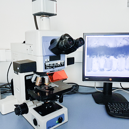
Nikon E800 is an upright wide-field fluorescence microscope intended for thin samples labelled with fluorescent or histological stains (excitation: CoolLED pE-300 W; emission: a few filter cubes; monochrome and color cameras available; deconvolution possible in Huygens). Polarization and DIC enable label-free imaging of samples in transmitted-light mode.
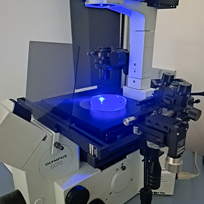
Olympus IX-70 is an inverted wide-field fluorescence microscope, ideal for fluorescence (excitation: mercury lamp; emission: a few filter cubes; camera currently unavailable) or bright-field imaging of larger specimens such as seedlings in Petri dishes, for counting cells, protoplasts or organelles in a counting chamber, and for direct observation in culture medium. The microscope is equipped with a micromanipulator.
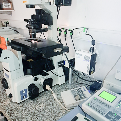
Nikon EZ-C1 is an inverted confocal microscope that has been in service for 20 years and remains in active use. Recommended for straightforward studies with up to three fluorophores, it works well with NIS-Elements deconvolution (wide-field fluorescence excitation: CoolLED pE-300 W, filter cubes, no camera; confocal imaging: 408, 488, 543 and 633 nm laser lines, filter cubes, 3 PMTs, transmitted light detector).
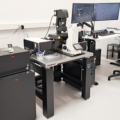
Leica Stellaris 8 Falcon is a state-of-the-art confocal microscope equipped with a white light laser and hybrid spectral detectors. It supports advanced techniques such as FRAP, FRET, laser micro-irradiation, spectral imaging, FLIM and FLIM-FRET. The system runs on LAS X software with the LIGHTNING module, 3D Viewer and AiviaGO, and is fitted with an Okolab environmental chamber for live-cell imaging.
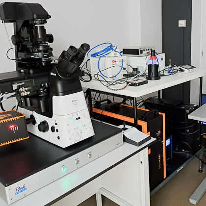
STEDYCON Abberior is a compact confocal system with a two-channel super-resolution (STED) mode, built on a Nikon Ti-E inverted microscope (no wide-field fluorescence). It is designed for imaging beyond the diffraction limit (excitation: 405, 488, 561 and 640 nm; STED depletion: 775 nm). The system is integrated with Huygens software and equipped with an Okolab environmental chamber.
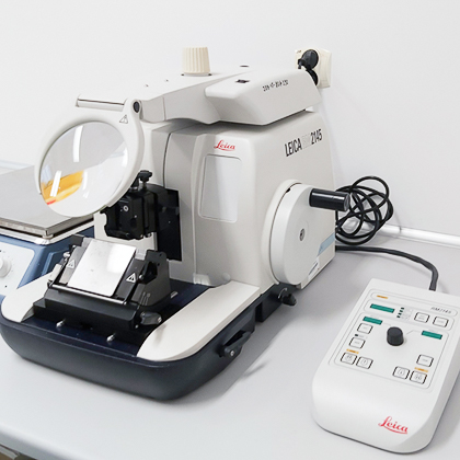
Leica RM2145 is a semi-motorized rotary microtome designed for sectioning paraffin-embedded specimens. Following immunocytochemical staining, thin preparations are ideally suited for observation using wide-field fluorescence microscopes.
Publications (IBB PAS affiliated)
Team
- Anna Anielska-Mazur, PhD, Employee, ORCID: 0000-0003-3679-7595